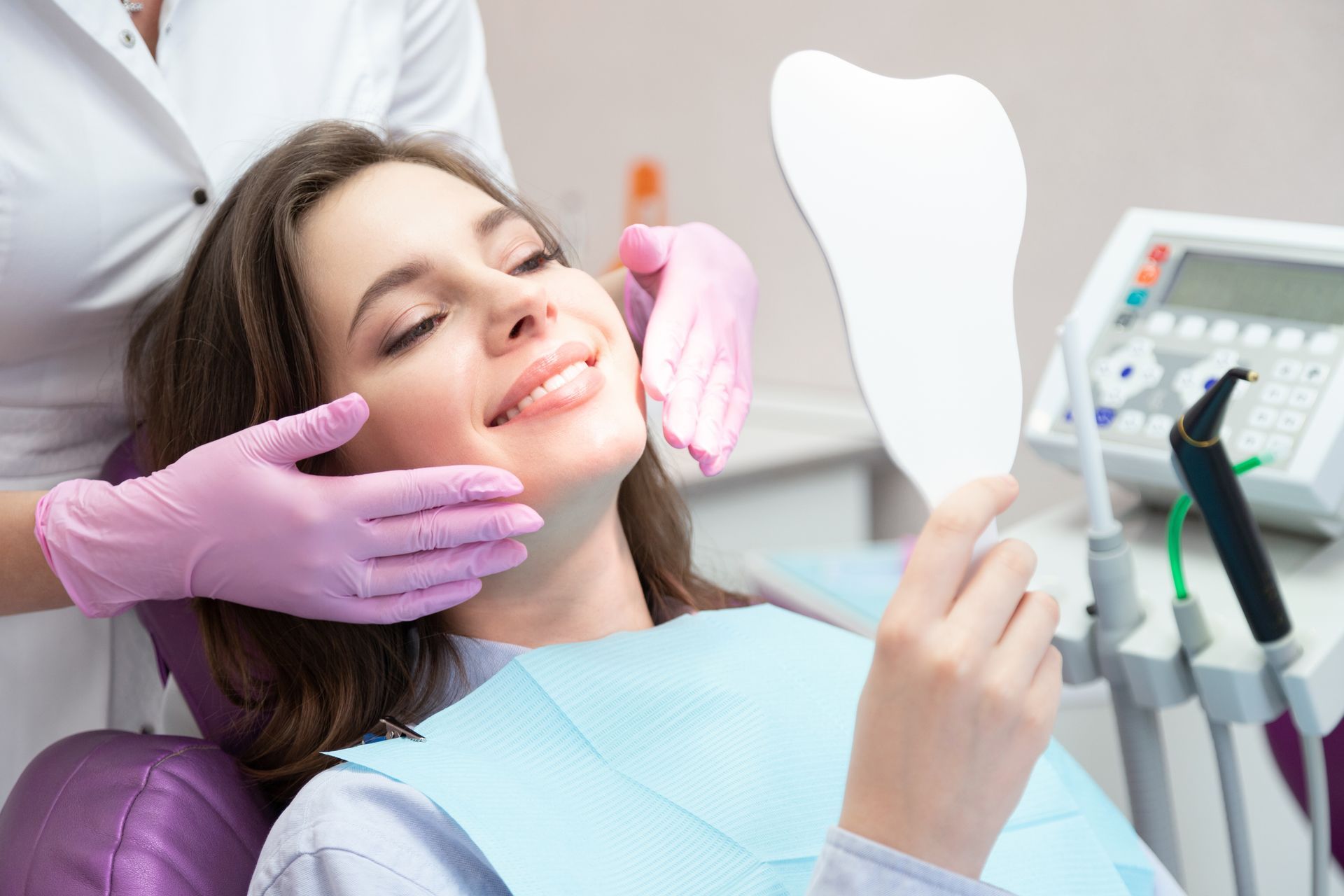
Discover 5 signs you may need cosmetic dentistry treatment in Phoenix, AZ. Desert Dental offers personalized care. Call today to schedule your visit.
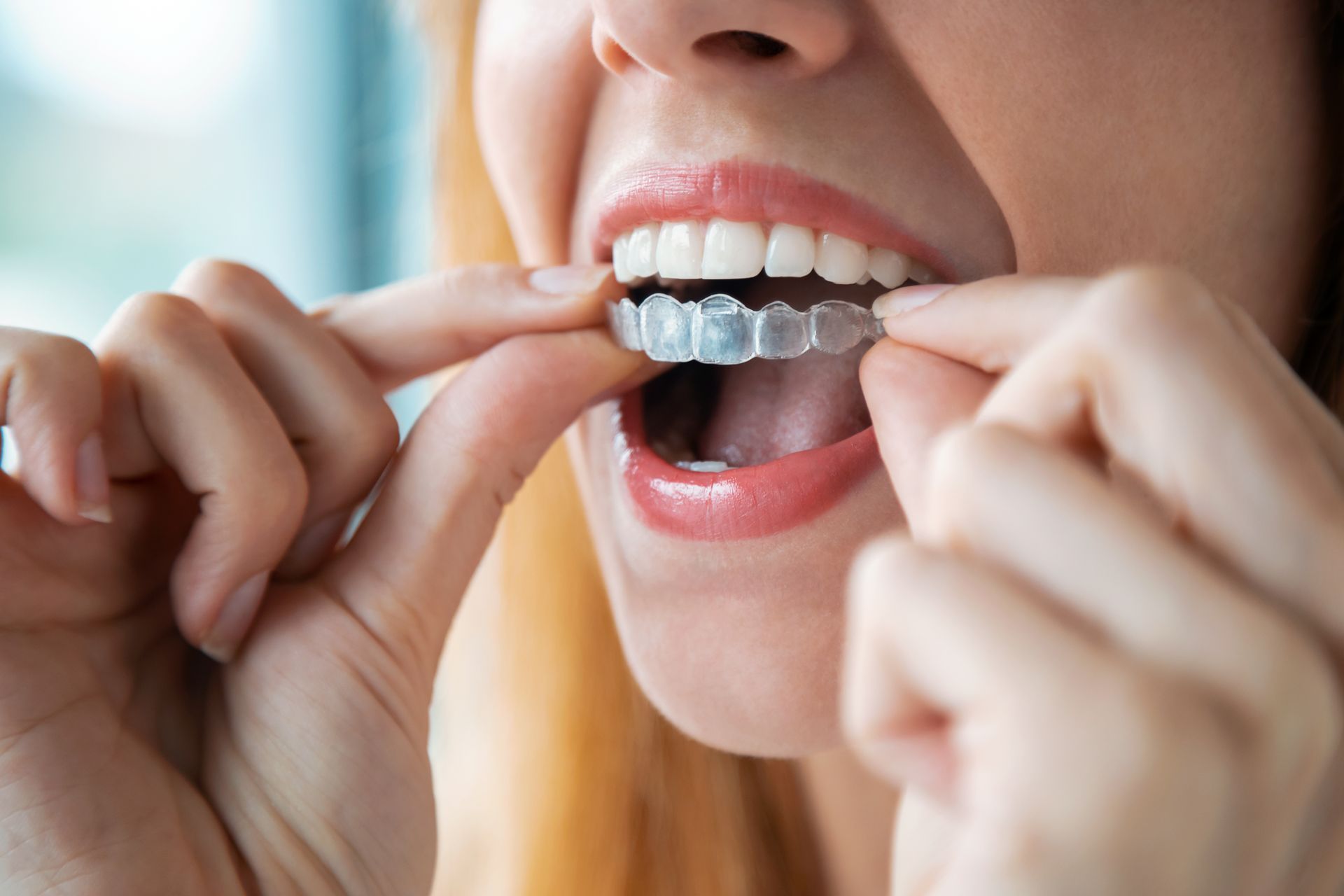
Active in Phoenix, AZ? Learn how Invisalign braces from Desert Dental fit your busy lifestyle. Click to read our blog and schedule your consultation now!
It's important to keep your dental health in check as you get older. To learn more about dental issues in the elderly, read our blog to learn more.
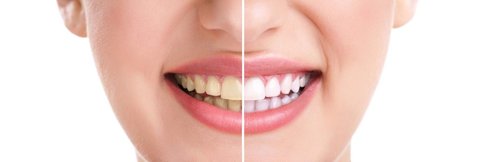
Do you want to try out teeth whitening? If so, you might have heard some misconceptions about them. Discover the truth behind three teeth whitening myths.

Hormonal imbalances can impact your oral wellness. Understand the delicate relationship between your mouth and the hormones your endocrine system produces.

When your teeth have damage, your dentist might recommend dental crowns. Before getting dental crowns, understand the following benefits of these fixtures.

Having bad breath all the time can be a sign of an underlying problem. Understand the possible culprits of bad breath to help you.

Gum contouring is a minor oral surgery procedure for adjusting the amount of gum tissue. Discover ways gum contouring enhances your smile.

Lumineers and veneers are effective options for people who want that Hollywood smile. Discover the differences between veneers and Lumineers.

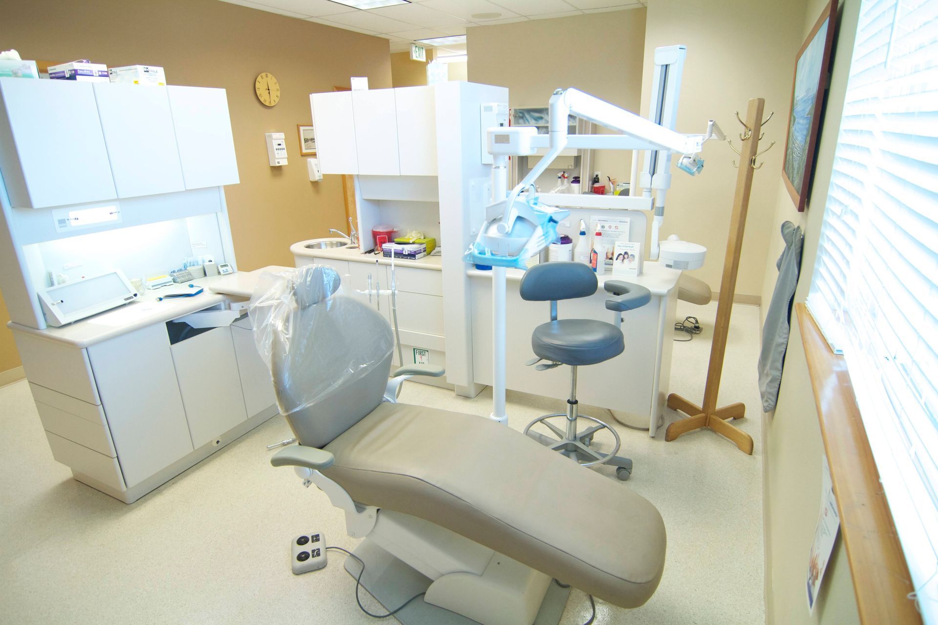
Many people dread dental appointments. Discover ways to ease your dental anxiety and protect your oral health with these helpful tips.